Home » Without Label » Anatomy Of Chest - 1 - A detailed knowledge of chest wall anatomy is crucial for reconstructions in this difficult patient population.
Anatomy Of Chest - 1 - A detailed knowledge of chest wall anatomy is crucial for reconstructions in this difficult patient population.
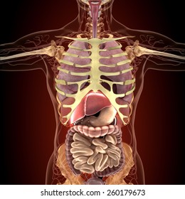
Anatomy Of Chest - 1 - A detailed knowledge of chest wall anatomy is crucial for reconstructions in this difficult patient population.. The chest wall is comprised of skin, fat, muscles, and the thoracic skeleton. 30 lines of the thoracic wall syllabus p. The muscles of the chest develop from the somites found in the mesoderm. Browse 6,407 chest anatomy stock photos and images available, or search for human anatomy to find more great stock photos and pictures. The chest is the area of origin for many of the body's systems as it houses organs such as the heart, esophagus, trachea, lungs, and thoracic diaphragm.
Anatomy of right side chest pain. An overview of the anatomy visible in a transverse computed axial tomographical image of the thorax (and part of the abdomen) performed with intravenous cont. Here, we break down the anatomy of your chest muscles. The pectoralis major and the pectoralis minor, known collectively as your pecs. The epidermis is the outermost layer that provides a protective, waterproof seal over the body.
Human Chest Anatomy Images Stock Photos Vectors Shutterstock from image.shutterstock.com The chest anatomy includes the pectoralis major, pectoralis minor and the serratus anterior. The first step in understanding thorax anatomy is to find out its boundaries. A typical heart is approximately the size of your fist: Table 1.1 lists the major anatomic structures within the thorax that are discussed. Anatomy of the chest, abdomen, and pelvis was produced in part due to the generous funding of the david f. In insects, crustaceans, and the extinct trilobites, the thorax is one of the three main divisions of the creature's body, each of which is in turn composed of multiple segments. An overview of the anatomy visible in a transverse computed axial tomographical image of the thorax (and part of the abdomen) performed with intravenous cont. Principal functions are the protection of internal viscera and an expandable cylinder facilitating variable gas flow into the lungs.
The circulatory system does most of its work.
The pectoralis major and the pectoralis minor, known collectively as your pecs. This section of the website will explain large and minute details of arterial anatomy of chest Knowledge of the anatomy of the whole cylinder (ribs, sternum, vertebra, diap … Chest muscles anatomy (1) pectoralis major muscle. The muscles of the chest develop from the somites found in the mesoderm. Chest a man's chest — like the rest of his body — is covered with skin that has two layers. Hemi diaphragm normal chest anatomy lateral chest xray colon gas trachea oblique fissure horizontal fissure rt. In insects, crustaceans, and the extinct trilobites, the thorax is one of the three main divisions of the creature's body, each of which is in turn composed of multiple segments. The chest is made up primarily of two muscles: Swensen fund for innovation in teaching. Basic thoracic anatomy and physiology an understanding of thoracic imaging requires knowledge of the anatomy being imaged, as described in this chapter, as well as the imaging techniques applied to the thorax, covered in chapter 2. An overview of the anatomy visible in a transverse computed axial tomographical image of the thorax (and part of the abdomen) performed with intravenous cont. Anatomy of right side chest pain.
Large, complex chest wall defects can be some of the most challenging problems a reconstructive surgeon must face, but successful outcomes may be reliably achieved by adhering to basic principles of adequate debridement followed by. The chest anatomy includes the pectoralis major, pectoralis minor and the serratus anterior. The anatomic illustrations are presented as… Chest a man's chest — like the rest of his body — is covered with skin that has two layers. The chest is made up primarily of two muscles:
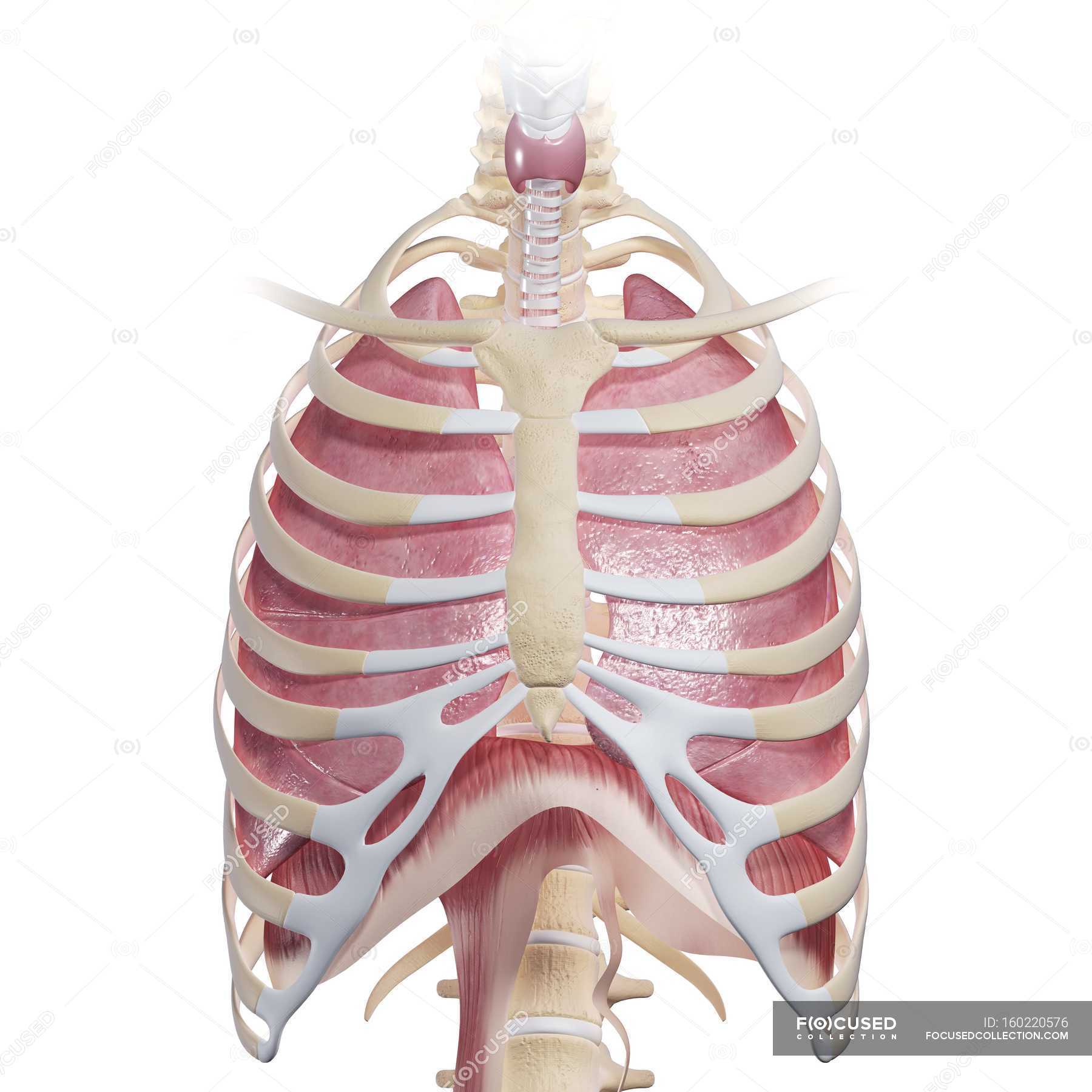
Human Chest Anatomy Larynx Physiology Stock Photo 160220576 from st.focusedcollection.com Chest a man's chest — like the rest of his body — is covered with skin that has two layers. The anatomic illustrations are presented as… The chest or thorax region of the upper body has a number of important organs that reside within it that may present with chest pain if they become compromised in. Anatomically, the heart is located in the anterior thoracic cavity; Chest muscles anatomy (1) pectoralis major muscle. 30 lines of the thoracic wall syllabus p. The chest is made up primarily of two muscles: The chest or thorax is the region between the neck and diaphragm that encloses organs, such as the heart, lungs, esophagus, trachea, and thoracic diaphragm.
Principal functions are the protection of internal viscera and an expandable cylinder facilitating variable gas flow into the lungs.
A detailed knowledge of chest wall anatomy is crucial for reconstructions in this difficult patient population. These myotomes divide into the epimere and the hypomere. Sternocleidomastoid muscle clavicle and ribs anatomy muscle anatomy chest sternocleidomastoid ribs anatomy chest muscles anatomy thorax rib muscles chest muscles chest anatomy illustration. 30 lines of the thoracic wall syllabus p. Chest a man's chest — like the rest of his body — is covered with skin that has two layers. Anatomy of the chest and shoulder, anatomy of the chest organs, anatomy of the chest wall, anatomy of the chest wall and pleura, anatomy of upper chest area, human. Large, complex chest wall defects can be some of the most challenging problems a reconstructive surgeon must face, but successful outcomes may be reliably achieved by adhering to basic principles of adequate debridement followed by. Three dimensional view of the female reproductive system, full frontal view. The circulatory system does most of its work. The chest or thorax region of the upper body has a number of important organs that reside within it that may present with chest pain if they become compromised in. Abdominal regions and organs 12 photos of the abdominal regions and organs 9 abdominal regions and its organs, abdominal cavity regions and organs, abdominal regions and associated organs, abdominal regions and its organs, abdominal regions and quadrants and organs, human anatomy, 9 abdominal regions and its. 12 cm (5 in) in length, 8 cm (3.5 in) wide, and 6 cm (2.5 in) in thickness. A good radiologist knows the anatomy because knowing where structures normally live and recognizing the location of an abnormality helps to make or narrow the differential diagnosis.
Abdominal regions and organs 12 photos of the abdominal regions and organs 9 abdominal regions and its organs, abdominal cavity regions and organs, abdominal regions and associated organs, abdominal regions and its organs, abdominal regions and quadrants and organs, human anatomy, 9 abdominal regions and its. Browse 6,407 chest anatomy stock photos and images available, or search for human anatomy to find more great stock photos and pictures. Here, we break down the anatomy of your chest muscles. Large, complex chest wall defects can be some of the most challenging problems a reconstructive surgeon must face, but successful outcomes may be reliably achieved by adhering to basic principles of adequate debridement followed by. The chest wall is comprised of skin, fat, muscles, and the thoracic skeleton.
Human Chest Anatomy Images Stock Photos Vectors Shutterstock from image.shutterstock.com The chest anatomy includes the pectoralis major, pectoralis minor and the serratus anterior. An overview of the anatomy visible in a transverse computed axial tomographical image of the thorax (and part of the abdomen) performed with intravenous cont. Radiology basics of chest ct anatomy with annotated coronal images and scrollable axial images to help medical students and junior doctors learning anatomy. 12 cm (5 in) in length, 8 cm (3.5 in) wide, and 6 cm (2.5 in) in thickness. Related posts of anatomy of the chest and stomach abdominal regions and organs. The chest wall is comprised of skin, fat, muscles, and the thoracic skeleton. These myotomes divide into the epimere and the hypomere. This chapter is an abbreviated review of thoracic anatomy as seen on chest radiographs and computed tomography (ct) of the chest.
System respiratory respiratory organs of human body digestive and respiratory system medical chest internal structure of human body medicine body lungs biology intestines stomach anatomy torso human internal.
In insects, crustaceans, and the extinct trilobites, the thorax is one of the three main divisions of the creature's body, each of which is in turn composed of multiple segments. Browse 6,407 chest anatomy stock photos and images available, or search for human anatomy to find more great stock photos and pictures. Abdominal regions and organs 12 photos of the abdominal regions and organs 9 abdominal regions and its organs, abdominal cavity regions and organs, abdominal regions and associated organs, abdominal regions and its organs, abdominal regions and quadrants and organs, human anatomy, 9 abdominal regions and its. The chest or thorax is the region between the neck and diaphragm that encloses organs, such as the heart, lungs, esophagus, trachea, and thoracic diaphragm. The first step in understanding thorax anatomy is to find out its boundaries. Large, complex chest wall defects can be some of the most challenging problems a reconstructive surgeon must face, but successful outcomes may be reliably achieved by adhering to basic principles of adequate debridement followed by. Anatomy of the chest and shoulder, anatomy of the chest organs, anatomy of the chest wall, anatomy of the chest wall and pleura, anatomy of upper chest area, human. Three dimensional view of the female reproductive system, full frontal view. An overview of the anatomy visible in a transverse computed axial tomographical image of the thorax (and part of the abdomen) performed with intravenous cont. Related posts of anatomy of the chest and stomach abdominal regions and organs. See chest anatomy stock video clips. It spreads out like a fan and covers the rib cage like an armor plate. The chest is the area of origin for many of the body's systems as it houses organs such as the heart, esophagus, trachea, lungs, and thoracic diaphragm.